


Nexcelom Cellometer Auto 2000 Cell Viability Counter
Description
Primary Cell Analysis: PBMCs, Stem Cells, and more
The Cellometer Auto 2000 is specifically optimized for analysis of primary cells from peripheral blood, cord blood, bone marrow, and other complex samples for use in a wide range of research areas, including:
- Nucleated Cells for Transplantation
- PBMCs for Immunology
- Splenocytes for Vaccine Development
- Stem Cells for Cellular Therapy
- Tumor Cell Suspensions for Oncology
Dual-color fluorescence allows for staining of live and dead nucleated cells, generating accurate viability results even in the presence of debris, platelets, and red blood cells. Accurate analysis of both 'messy' and 'clean' samples enables the Auto 2000 to evaluate samples at a variety of points throughout sample processing - from initial collection, to separation, to cryopreservation.
No Interference from Red Blood Cells, Platelets, or Debris
The dual-fluorescence AO/PI method utilizes nuclear staining dyes that bind to nucleic acids in the cell nucleus. Because most mature mammalian red blood cells do not contain nuclei, only live and dead mononuclear cells produce a fluorescent signal. There is no need to lyse red blood cells, saving time and eliminating an extra sample preparation step. Red blood cells, platelets, and debris are not counted in the fluorescent channels.
| Sample | Measurement | Total nucleated | All | RBC | % RBC | n |
|---|---|---|---|---|---|---|
| PBMC+RBC | Mean | 1.26E+07 | 1.39E+07 | 1.23E+06 | 8.9% | 10 |
| CV | 6.2% | 8.8% | ||||
| PBMC+1/2RBC | Mean | 1.21E+07 | 1.27E+07 | 5.82E+05 | 4.6% | 10 |
| CV | 4.8% | 5.4% | ||||
| PBMC+1/4RBC | Mean | 1.22E+07 | 1.24E+07 | 2.27E+05 | 1.8% | 10 |
| CV | 7.9% | 7.3% |
Fresh human PBMCs (peripheral blood mononuclear cells) were spiked with varying amounts of RBCs (red blood cells.) All cells (nucleated + RBC) were counted in the bright field channel. Nucleated cells were then counted in the green fluorescent channel. Varying amounts of RBCs (1.8%, 4.6%, and 8.9%) did not affect the nucleated cell count.
The advantage of fluorescent counting for primary cells
The images below demonstrate the advantage of fluorescent counting for primary cells. The bright field image shows the combination of nucleated cells, red blood cells, and platelets present in the sample. Only the live and dead nucleated cells are visualized and counted in the green and red fluorescent channels.
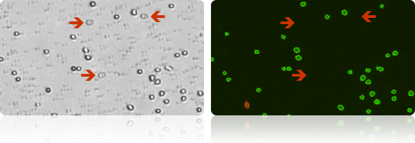
Several red blood cells are indicated in the bright field image (above, left). The red blood cells are not visible in the fluorescent image (above, right) detecting cells stained with nuclear staining dye.
Analysis of Clumpy and Irregular-shaped Cells
Including NCI-60 and clumpy MCF-7 Cells
NCI-60 is a group of 59 human cancer cell lines (originally 60) developed by the National Cancer Institute for screening purposes.
- 57% of the NCI-60 cell lines are clumpy, contain debris, or display large variations in cell shape or size
- All 59 NCI-60 cell lines have been successfully validated on the Cellometer Cell Counter
All 40 of the NCI Comprehensive Cancer Centers use Cellometer Cell Counters.
Clumpy Cells
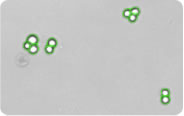 The MCF-7 breast cancer cell line can be very clumpy. The Cellometer pattern-recognition software identifies and counts individual cells within these cell clumps for accurate analysis (shown above).
Irregular-shaped Cells
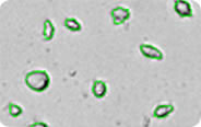 The Cellometer cell roundness setting can be adjusted for recognition and counting of irregular-shaped cells, such as RD cells and activated T-cells.
Features
Features & Benefits of Cellometer Auto 2000
- 30 Second Cell Concentration & Viability
- User-Friendly Touch Screen
- Primary Cell Analysis
- Live/Dead Nucleated Cell Counts
- No Interference from Red Blood Cells
- Cell Images for Data Verification
- Analysis of Clumpy and Irregular-shaped Cells
- Cell Size Analysis & Size-based Counting
- 10x Faster than Manual Counting
- No Washing or Contamination
- Auto-Save Data and Cell Images
- Dedicated On-Line and On-Site Support
The Auto 2000 Allows Users to:
- Increase throughput
- Increase accuracy
- Improve consistency
- Ensure all data is correctly captured
- Count difficult cells (clumpy, irregular-shaped)
- Eliminate judgment errors, miscounts, interference from red blood cells and user-to-user variability
User Friendly Touch Screen
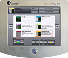 The user-friendly touch screen displays pre-optimized assays for common cell types. User-specific protocols can be easily created and saved to the menu. The simple, pre-optimized assays make the instrument easy to operate and simplify training for new users.
With the click of a button, researchers can select a new assay with saved counting parameters, making the instrument ideal for labs testing a variety of cell types. Auto-save options allow users to quickly test a series of samples.